

The New England Journal of Medicine is the world’s leading medical journal and website.
- Tweets 38,355
- Following 432
- Followers 1,037,829
- Likes 2,243


ALT Graphs from the Original Article "Atorvastatin, Cardiovascular Events, and Disability-free Survival in Older Adults." The trial name, STAREE, is in parentheses after the title. The graphs show the cumulative incidence of primary end-point events (death from cardiovascular causes, nonfatal myocardial infarction or stroke, or coronary revascularization, whichever occurred first). The trial results are being presented at the European Society of Cardiology Congress 2026. The logo of the New England Journal of Medicine is at the bottom.

ALT This image contains four graphs. Panels A and B show progression-free survival and overall survival among all patients who received anitocabtagene autoleucel (anito-cel), a B-cell maturation antigen (BCMA)–directed autologous chimeric antigen receptor (CAR) T-cell therapy with a synthetic D-domain binder (ddBCMA). Panels C and D show progression-free survival among patients according to the presence or absence of extramedullary disease (EMD) and according to the β2-microglobulin (β2M) level (≥5.5 or <5.5 mg per liter), respectively.

ALT This image contains five graphs. Panel A shows anito-cel vector transgene levels in peripheral blood per microgram of genomic DNA (gDNA). Panels B–E show changes in serum soluble BCMA levels and in concentrations of CAR T-cell activation–associated serum analytes interferon-γ, interleukin-2, and interleukin-6 that were evaluated at baseline and after the anito-cel infusion.
![This image contains four figures. Panel A shows the size and structure of the dual variable heavy-chain domain of a heavy chain–only antibody (dual-VHH) and ddBCMA binders and biolayer interferometry binding data demonstrating strong avidity effects for the dual-VHH binder, whereas no avidity effect was observed with the ddBCMA binder. Panel B shows a schematic of the research-grade CAR constructs expressed in T cells and used in subsequent experiments. Panel C shows immune synapse formation between ddBCMA CAR or dual-VHH CAR T cells and BCMA-expressing K562 cells as assessed by imaging flow cytometry. Panel D shows cytotoxicity (the percentage of tumor cells killed) and cytokine release values (granulocyte–macrophage colony-stimulating factor [GM-CSF], interleukin-2, and interferon-γ) with primary bone marrow mononuclear cells from patients with multiple myeloma after coculture with ddBCMA CAR T cells or dual-VHH CAR T cells, with technical replicates shown as means.](/pic/orig/media%2FHTKie1DXYAEWJJL.jpg)
ALT This image contains four figures. Panel A shows the size and structure of the dual variable heavy-chain domain of a heavy chain–only antibody (dual-VHH) and ddBCMA binders and biolayer interferometry binding data demonstrating strong avidity effects for the dual-VHH binder, whereas no avidity effect was observed with the ddBCMA binder. Panel B shows a schematic of the research-grade CAR constructs expressed in T cells and used in subsequent experiments. Panel C shows immune synapse formation between ddBCMA CAR or dual-VHH CAR T cells and BCMA-expressing K562 cells as assessed by imaging flow cytometry. Panel D shows cytotoxicity (the percentage of tumor cells killed) and cytokine release values (granulocyte–macrophage colony-stimulating factor [GM-CSF], interleukin-2, and interferon-γ) with primary bone marrow mononuclear cells from patients with multiple myeloma after coculture with ddBCMA CAR T cells or dual-VHH CAR T cells, with technical replicates shown as means.

ALT A quote from a Perspective published in the New England Journal of Medicine reads as follows: "I want to do something, but all I can do is feel but an ounce of the powerlessness you must feel, so I leave. I write my note: tidy all you are within the confines of S.O.A.P." The Perspective is titled "Gaps and Borders" and the author is Brandon Oddo, B.S. The background color is teal and includes the NEJM logo.
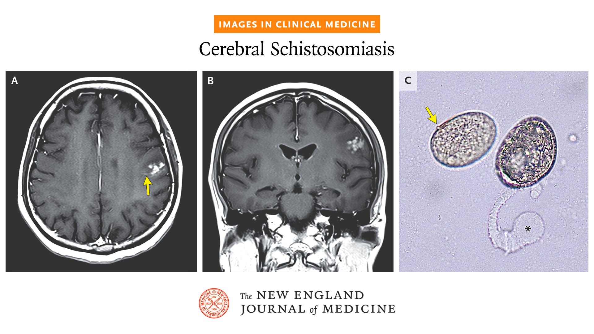
ALT Three medical images from the Images in Clinical Medicine article "Cerebral Schistosomiasis." Panels A and B: Axial and coronal T1-weighted images obtained after administration of contrast material show an area of linear enhancement (arrow) associated with a cluster of enhancing nodules and surrounding edema in the left frontal lobe. Panel C: A sample from a circumoval precipitin test for Schistosoma japonicum — which involves incubation of live S. japonicum eggs with patient serum — that was performed. The NEJM identity sits at the bottom.

ALT Timeline of a clinical course highlighting the treatment and progression of severe Oya virus infection in a patient who also had hepatitis, enteritis, and signs of coagulopathy. It tracks hospital admissions, symptoms, treatments, and recovery progress. Key details include initial symptoms like fever and chills, transfer to Guangzhou Eighth People's Hospital, various treatments such as ibuprofen, antibiotics, antivirals, and plasma transfusion, and the resolution of symptoms. The chart ends with the patient's discharge and follow-up.

ALT An image announcing a new audio interview with Christopher B. Forrest, M.D., Ph.D., on Developmental Ecosystems and Health. There is a photo of Dr. Forrest over a decorative soundwave graphic.

ALT A quote from an NEJM Voices blog post reads as follows: “When we show up, we show up as a friend, not as a clinician — being a clinician lets us be the friend who does not need to make it about themselves.” The author is Theodore Iwashyna, M.D. The background color is teal and includes the NEJM Voices logo.


ALT A illustration showing anticoagulation for atrial fibrillation with intermediate stroke risk. On the left, a medicine box labeled "Anticoagulant Tablets" with two pills is shown. In the center, a silhouette of a patient with a highlighted heart. Above, a round inset displays a heart rate monitor graph representing atrial fibrillation. To the right, a brain is depicted with an inset showing a blood vessel and a warning symbol, representing intermediate stroke risk.
![Four graphs show the summary of efficacy end points up to week 12. Panel A shows the values on the Maintenance of Wakefulness Test (MWT), which range from 0 to 40 minutes (normal ability to stay awake, ≥20 [black dashed line]). Panel B shows the total scores on the Epworth Sleepiness Scale (ESS), which range from 0 to 24, with higher scores indicating greater daytime sleepiness (normal, ≤10 [black dashed line]). Panel C shows the weekly cataplexy rate, which includes both partial and full cataplexy events. Panel D shows the total scores on the Narcolepsy Severity Scale for Clinical Trials (NSS-CT), which range from 0 to 57, with scores of 0 to 14 indicating mild narcolepsy symptoms, 15 to 28 moderate symptoms, 29 to 42 severe symptoms, and 43 to 57 very severe symptoms; the black dashed lines indicate the thresholds for the different levels of symptom severity.](/pic/orig/media%2FHTFY3G3WkAAkKY_.png)
ALT Four graphs show the summary of efficacy end points up to week 12. Panel A shows the values on the Maintenance of Wakefulness Test (MWT), which range from 0 to 40 minutes (normal ability to stay awake, ≥20 [black dashed line]). Panel B shows the total scores on the Epworth Sleepiness Scale (ESS), which range from 0 to 24, with higher scores indicating greater daytime sleepiness (normal, ≤10 [black dashed line]). Panel C shows the weekly cataplexy rate, which includes both partial and full cataplexy events. Panel D shows the total scores on the Narcolepsy Severity Scale for Clinical Trials (NSS-CT), which range from 0 to 57, with scores of 0 to 14 indicating mild narcolepsy symptoms, 15 to 28 moderate symptoms, 29 to 42 severe symptoms, and 43 to 57 very severe symptoms; the black dashed lines indicate the thresholds for the different levels of symptom severity.

ALT A quote from a Perspective published in the New England Journal of Medicine reads as follows: "Signs of another pendulum swing in Americans’ relationship with biomedical science and with scientific and public health elites may already be emerging." The Perspective is titled "The End of the War on Biomedical Science?" and the authors are David Blumenthal, M.D., M.P.P., and James A. Morone, Ph.D. The background color is orange and includes the NEJM identity.

ALT CT scan of a neck showing diffusely enlarged cervical lymph nodes on the left side with perinodal fat stranding, as well as a lymph node at the level of the lower jugular with internal heterogeneity. Text reads, "Case Challenge" and "Cast your vote before October 8."

ALT A graph showing the cumulative incidence of hematologic complications in patients with Shwachman–Diamond syndrome, including myelodysplastic syndromes, acute myeloid leukemia, severe bone marrow failure, and hematopoietic stem-cell transplantation indications.

ALT A table showing the evaluation of the effectiveness of lung cancer screening methods. The first column lists screening method, including low‑dose computed tomography and chest radiography with or without cytologic examination of sputum. For each method, there is body of evidence reviewed and classification for reduction in lung cancer mortality and incidence of stage III or IV lung cancer.

ALT A table showing the role of emerging tools to enhance lung cancer screening. It lists various tools and their roles, including AI-assisted image reading, molecular biomarkers, and virtual shared decision-making tools. Columns indicate specific functions such as assist with screening uptake and tailor screening interval, with "Yes" or "No" answers.

ALT This image consists of CT images of the chest, abdomen, and pelvis. Panel A: A contrast-enhanced image of the chest shows a stable pulmonary nodule in the right middle lobe (circle), a finding compatible with known pulmonary metastatic disease. Panel B: A contrast-enhanced image of the abdomen and pelvis shows decreased attenuation throughout the liver (arrow) as compared with the spleen (asterisk), a finding compatible with hepatic steatosis.

ALT This image consists of initial MRI of the head. A T2-weighted fluid-attenuated inversion recovery (FLAIR) image (Panel A), a diffusion-weighted image (Panel B), and a T1-weighted contrast-enhanced image (Panel C) show a region of encephalomalacia in the posterior aspect of the left temporal lobe and left occipital lobe (arrows) with surrounding gliosis (Panel A, asterisks), findings compatible with a chronic infarct.

ALT This image consists of repeat MRI of the head. Axial T1-weighted images obtained before and after the administration of contrast material (Panels A and B, respectively) show marked enhancement involving the mammillary bodies on both sides (arrows). T1-weighted contrast-enhanced images in the coronal and sagittal planes (Panel C and D, respectively) also show avid enhancement involving the mammillary bodies (arrows), which can be seen anterior to the midbrain (Panel D, asterisk). Axial T2-weighted FLAIR images at the level of the midbrain and thalami (Panels E and F, respectively) show no definitive signal abnormality in the periaqueductal gray matter (Panel E, circle) or medial thalami (Panel F, arrows).

ALT A graph showing the cumulative incidence of primary end-point events (intention-to-treat population).

ALT A quote from a Perspective published in the New England Journal of Medicine reads as follows: “Decades of design choices have led to systems in which markets are the primary drivers of health, caregiving infrastructure is fragmented, and prevention is devalued and chronically underfunded.” The Perspective is titled “Declining Child Health in the United States — Addressing the Developmental Ecosystem” and the authors are Christopher B. Forrest, M.D., Ph.D., and Neal Halfon, M.D., M.P.H. To the right is an illustration of an iceberg representing factors driving declines in child health. The iceberg is separated into four sections: "Surface Level: Declines in physical, neurodevelopmental, and mental health"; "Proximal Drivers: Developmental processes altered by exposures throughout the life course"; "Structural Drivers: Degraded natural and social systems"; and "Foundational Drivers: Antiquated cultural narratives about children’s needs."

ALT Two anteroposterior radiographs of the patient's legs from "Case Records of the Massachusetts General Hospital: A 4-Year-Old Boy with Fatigue, Imbalance, and Frequent Falls."

ALT Cover page of NEJM.org dated September 24, 2026. It includes sections like Specialties, Topics, Multimedia, Current Issue, and Learning/CME. Featured content includes "Narcolepsy Research Recognized through the Lasker Award" and an audio interview with Christopher Forrest on Developmental Ecosystems and Health. The page also highlights the Images in Clinical Medicine article "Cerebral Schistosomiasis," the Quick Take "Anticoagulation in Atrial Fibrillation with Intermediate Stroke Risk," and the editorial "Science behind the Study: CAR with Synthetic Antigen-Binding Domain for Myeloma."

ALT A quote from an NEJM Voices blog post reads as follows: “We must have the freedom, as physicians, to tell patients what they need to hear rather than what they want to hear.” The author is Michelle Kittleson, M.D. The background color is red and includes the NEJM Voices logo.

ALT A table showing the evaluation of the effectiveness of lung cancer screening methods. The first column lists screening method, including low‑dose computed tomography and chest radiography with or without cytologic examination of sputum. For each method, there is body of evidence reviewed and classification for reduction in lung cancer mortality and incidence of stage III or IV lung cancer.