

Cortical circuits lab @ NYU CNS. Lasers, neurons, coffee.
NYU CNS
Based in United States
- Tweets 541
- Following 415
- Followers 586
- Likes 185

7/ When she targeted the C2 barrel in vS1, she found that it reduced touch responsiveness in vS2 in a whisker-specific manner.

1
1
204
6/ She then lesioned the 'touch patch’ in vS2, and found that this diminished touch responses in vS1.

1
1
90
5/ She found that despite their rarity, multiwhisker cells carried a large share of touch activity in both directions.

1
1
91
4/ Lauren then used retrograde AAV injections in both areas, targeting just the portion of each area that responded to a specific whisker, and asked how somatotopically-matched regions communicated.

1
1
104
3/ Despite making up only ~1% of neurons, multiwhisker cells nevertheless carried a large fraction of the touch response superficially.

1
1
105
2/ Lauren first imaged L2-4 in both whisker S1 and S2 in mice with two spared whiskers. In both areas, neurons in deeper L2/3 and L4 were mostly responsive to single whiskers, whereas superficial neurons were broadly tuned and responded to multiple whiskers.

1
1
173

Broadly tuned cells also showed greater stability in terms of the correlation of touch responsiveness, measured using an encoding model, across days.

1
1
121
Cells with broader tuning on day 1 – multiwhisker or bidirectional single whisker neurons – were consistently more touch responsive.

1
1
137
Alisha studied drift using a two-whisker touch task, classifying neurons responsive to different combinations of forward and backward touches by both whiskers.

1
1
233
We propose that these rare multiwhisker neurons with motor-related activity may act as key nodes for sensorimotor integration, potentially bumping motor ensembles to threshold as sensory evidence in the form of touch is gathered by the animal.

125
Finally, he showed that multiwhisker neurons, especially those with strong responses, were unusually likely to also participate in licking or whisking ensembles.

1
124
He also looked at long term stability, positing that neurons involved in sensorimotor integration would especially benefit from having more stable activity. Indeed, the most responsive 25% of neurons were far more stable than weakly responsive neurons.

1
26
Ravi then imaged animals with retrograde tracer injections into vS1 or vM1. For multiwhisker touch, whisking, and licking neurons, the most responsive 25% of neurons were far more likely to project across areas than the least responsive cells.

1
30
In both vS1 and vM1, Ravi finds that touch activity is disproportionately confined to multiwhisker neurons, especially the most responsive 25% of multiwhisker touch cells.

1
48


6/ To ask how these areas influence one another, we used columnar-scale lesions to ablate the portion of either vS1 or vS2 responsive to the two spared whiskers.

1
180
5/ Despite being a minority of in both areas, broadly tuned neurons turn out to be the largest contributor to touch activity relayed in both directions.

1
155
4/ Lauren then used small injections of retrograde tracer into patches of cortex responsive to the spared two whiskers and imaged the activity conveyed from somatotopically matched regions in the other area.

1
165
3/ In both vS1 and vS2, she finds that broadly tuned neurons – those responding to two whiskers – become more common as information flows from L4 to L2. They also end up carrying most of the touch response.

1
167
2/ Lauren began by imaging the parts of mouse vibrissal S1 and S2 that represent the same whiskers

1
266
Can this effect be enhanced? Ravi trained mice on a combined optical microstimulation / whisker touch task, and after 10 days of this, the overlap between the representations increased substantially.

1
167
Using optical microstimulation and 2p imaging of barrel cortex, Ravi finds that touch neurons do indeed overlap disproportionately with the microstimulation responsive population, whereas whisking neurons do not. Opsin+ neurons again showed a weaker effect than Opsin- neurons.

1
177
In the model, directly stimulated ('Opsin+') neurons showed a weaker effect than indirectly stimulated ('Opsin-') neurons, suggesting that over successive cortical processing stages, the evoked activity becomes ever more natural.

1
1
171
Ravi first explored this in a model of mouse barrel cortex with a subpopulation of neurons having elevated recurrence. Simulated random optogenetic stimulation in the model disproportionately engaged this recurrently coupled subnetwork.

1
1
277
(5/7) Repeatedly exposing animals to paired optogenetic and touch stimuli increased overlap between photoresponsive and touch populations.

1
1
(4/7) We found that photostimulation engaged (or “overlapped with”) touch neurons more than expected by chance.

1
(3/7) We used optogenetics to stimulate excitatory neurons in mouse primary vibrissal somatosensory cortex. We then compared photostimulus-evoked activity (🔦⚡️) with natural activity evoked by whisker touch and movement (🐭).

1

Finally, we found that the degree of sparsification predicted the rate of learning: mice that had the most stable photoresponsive populations learned the fastest. (11/13)

1
We found that photoresponsive neurons increasingly overlapped with neurons responding to whisker touch and movement. (9/13)

1
1
After this initial sparsification stabilized, the responsive population was still dynamic and showed a steady rate of neural turnover or “representational drift”. (6/13)

1
1
Throughout, we recorded neural activity using two-photon calcium imaging. We saw rapid sparsification of the photoresponsive population. (5/13)

1
1
We used a miniature LED to photostimulate opsin-expressing neurons in layer 2/3 of barrel cortex and asked mice to tell us how many light pulses were delivered on each trial. (4/13)

1
1
Neuroscientists have been generating artificial percepts using direct cortical stimulation for decades. Perhaps most famously, Penfield and colleagues found they could evoke sensations when electrically stimulating somatosensory cortex. (2/13)

1
1

Espresso machine is back after power spike toasted the control board. No better way to start a Saturday.

1
11

Sadly, here is the updated version; NYC releases numbers twice daily, I think you got the AM release ... man you had me hopeful :(

2
2

Self-isolation day 2: I got some gymnastics rings to do some weight training while not being able to hit the gym.
My daughter clearly has a much better use for them.

2
This gives me flashbacks to my PhD days where, for an entire summer, an undergrad would come in, click on ommatidia for 6-8 hours, and go home.
Locusts have way too many ommatidia (~7500!).


6/ Ablation effect depends on how much of the touch representation we degrade (black: silent; green: whisking; blue: touch)

1
1
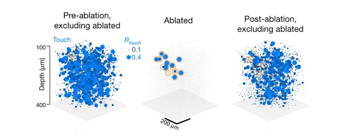
4/ We then use cellular-resolution ablation in mice following volumetric imaging to see if touch and whisking representations in barrel cortex show this decline

1
3/ The model makes a very simple prediction: if elevated recurrence is present in a subnetwork, the network should be highly sensitive to removing a handful (20 neurons of 200) of its members - response in such networks will decline.

1
2/ Longstanding theoretical and experimental work argues that recurrent excitatory circuitry in cortex mediates amplification; we begin by building a model that confirms this intuition:

1
2/2 Tanke is much more thorough; they pre-characterized cells based on if they were touch responsive. All 6 (because touch cells are rare) are above the diagonal, so in principle can drive responses (though 3/6 are likely NS).

1
1/2 Look at Fig 3 (esp. e); top 3 cells are FS cells, and only 1 non-FS cell breaks from the trend. So basically 1 cell tops.

2
Two weeks without our machine, first figuring out what was making it explode in the middle of the night, then waiting for parts.
Today she's back!
Yes, I need to up my latte art game.

4
